Operating microscope in Endodontics (Root canal treatment)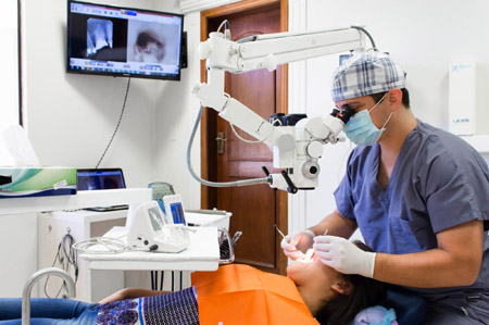
The practice of endodontics requires precision and big attention to detail. These depend on the training, abilities, and experience of the physician. The majority of endodontic procedures are done in confined and dark spaces, and a fraction of a millimeter can determine the result of the treatment. In the past decades, endodontics has gained not only basic and clinical scientific knowledge, but also has benefited from huge technological advances. Due to the intricate nature of endodontic treatment, professionals have always seeked to improve the view of the operative field or work area.
Advantages of Dental Microscopes
A better visualization requires improved magnification and illuminations, and the microscopes, as well as the loups, have been widely adopted. The dental microscopes possess a variety of advantages compared to the loups. Because the microscopes are units with their own support, the increase of lenses and prisms is not an inconvenience. This has significant implications regarding ergonomics and visualization.
Microscopes provide an adjustable magnification in a range of approximately 2x to 25x, while most loups provide a fixed magnification between 2,5x and 6x. The magnification can be divided into low (~2x-8x), medium (~8x-16x), and high (~16x-25x). The low, medium, and high magnifications are applicable to the different stages of procedures in surgical and not surgical endodontic treatments.
The use of a microscope increases significatively the operator’s precision.
Uses of the Microscope in Non-Surgical Procedures: For the endodontics specialist, the dental microscope is useful for the diagnosis and clinical procedures. During the diagnosis, the microscope can help in identifying caries, insufficient margins of restorations and crowns, or in the evaluation of fissures or fracture lines. During root canal therapy, the magnification and illumination provided by the operating microscope helps in the removal of caries, access preparation, removal of calcifications, identification of main root canal and accessories, identification of fissures and fracture lines, and in the internal resorption treatment. Under the microscope, subtle color and texture changes become apparent, allowing the professional to perform the procedures with more security.
High magnification can help in the localization and instrumentation of obstructed and calcified roots, in the identification of bifurcations, in the removal of obstructions such as calcifications, in the evaluation of the correct preparation and removal of remaining pulp tissue and root filling.
Increased visualization also helps in the treatment of dental anomalies, such as fused teeth or dens in dente, among others. In the re-treatments, the microscope is useful in the identification and removal of filling materials such as sealant, pastes or gutta-percha, silver cones, bolts, or fracture instruments. It also helps in the non-surgical reparation of perforations, allowing the cleaning and placement of the reparation material to be done with more precision.
Uses of the Microscope in Surgical Procedures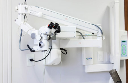
Surgical endodontics has been completely transformed by the microscopic procedures. The incorporation of the microscope, alongside the use of ultrasonic tips and biocompatible filling materials, has evolved the classic apicoectomy into the modern endodontic microsurgery. All of the steps in endodontic microsurgery are carried out under different degrees of magnification, including the flap preparation, osteotomy, identification of root apices, resection of the root end, removal of inflammatory tissue, observation of the sectioned surface, preparation and filling of the root end, and suture. The microscope is also useful for the reparation of cervical or external reabsorptions and of perforations.
Conclusion
The dental operating microscope has become an integral part of the practice of endodontics.
For surgical and non-surgical endodontic therapy, excellence is essential. Besides the obvious benefits for the clinical practice, literature demonstrates the increase of success rates in endodontic treatments (surgical and non-surgical) when done with the microscope, in comparison to the ones done with loups or without magnification.
The treatments carried out with the use of the operating dental microscope result in superior care for the patients, and modern endodontic therapy is more effective due to its use.
Electronic Foramen Locator in Endodontics (Root canal Treatment)
The presence of microorganisms inside the root canal system is generally accepted as the main cause of the failure of endodontic treatments, also known as root canal treatment. Thus, maximum cleaning and disinfection, and filling until the precise limit of the root canal are fundamental. Moreover, apical constriction (fig. 1) is considered by general rule as the anatomical reference point, until where the instrumentation and filling must reach. However, locating the apical constriction, from a clinical perspective, is very hard, due to the fact that its position and formation are highly variable.
Working length
The distance between the coronal reference point and the apical constriction is known in endodontics as “working length”. The methods to determine the working length include tactile sensibility, the knowledge of the different canals’ lengths and their anatomy, the evaluation of X-rays prior to the procedure, and the electronic foramen locators. Traditionally, X-rays have been the most used method to obtain information on the anatomy of the root canal and surrounding tissue.
The correct determination of the working length is fundamental for the complete cleaning, disinfection, and filling of the root canal, as well as for the prevention of mistakes in the procedure that may affect the final result of the treatment.
Disadvantages of the traditional X-ray method
The measurement of the working length performed by X-ray has several limitations:
- Exposure to radiation,
- Time spent, and
- difficulty in interpretation because it is a two-dimensional image that is often superimposed with other anatomical structures and is subject to the observer’s interpretation.
Electronic Foramen Locator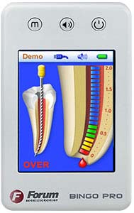
Foraminal electronic locators are electronic devices used in endodontics to determine the position of the apical constriction and thus determine the length of the root canal (working length). The end of the root canal or apical constriction has a specific resistance to electrical current, and this is measured using a pair of electrodes, which are usually placed on the patient’s lip and on the endodontic instrument. The first devices worked with the principle of resistance to the current. The problem with these devices was that conductive fluids, such as blood, exudate, or irrigators within the duct allowed for current flow and therefore false readings. The new devices are based on impedance and use alternating currents of two frequencies; these measure and compare two electrical impedances that change as the file progresses towards the end of the conduct. The readings of these devices are much less affected by conductive fluids present in the ducts and have demonstrated from 80 to 95% accuracy in the identification of the apical foramen.
Advantages of Electronic Apical Locators
- The measurement is more accurate and reliable than with X-ray methods.
- The procedure is quick and easy
- They can be used repeatedly at different stages of treatment
- Decreased amount of exposure to radiation to both patient and medical personnel
- Perforations in the root canal can be detected
- It is a method to find the physiological foramen, not only the radiographic apex
- They allow the use of any type of file
- They are able to take wet ducts measurements
- They can be a method to determine the level of horizontal fractures
Limitations of Electronic Apical Locators
- The use of Electronic Foraminal Locators is CONTRAINDICATED in patients with PACEMAKERS
- Their use demands careful technique and requires operator training and experience.
Observations
- A preliminary diagnostic x-ray and a final x-ray are always necessary for the evaluation of treatment results. Periodic monitoring X-rays are essential for the evaluation of the results and their frequency depends on each case and should be indicated by the specialist.
- Whenever x-rays are taken the patient should be protected by a thyroid vest and collar.